Ira Tabas, Ph.D., is interested in understanding more about how our bodies clear dead cells. The Columbia University professor spoke Dec. 4 as part of the NIEHS Distinguished Lecture Series.
“Dr. Tabas did an outstanding job weaving together a fascinating story and doing so in a disarmingly clear and simple manner,” said Michael Fessler, M.D., who hosted the talk by Tabas.
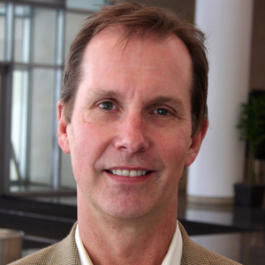
 Tabas is the Richard J. Stock Professor and Vice-Chair of Research in the Department of Medicine at Columbia University. (Photo courtesy of Steve McCaw)
Breaking down the garbage
Tabas described current knowledge that links mitochondria, which are structures that produce most of a cell’s energy, to a process for disposing of multiple dead cells, known as efferocytosis. Defects in this removal process can lead to autoimmune disease, chronic lung disease, neurodegenerative disease, atherosclerosis, and other diseases.
Efferocytosis requires cells called macrophages to remove the dead cells using a mechanism known as phagocytosis, or cell eating. To do this, the macrophage has to dramatically rearrange its cellular membrane and handle a large metabolic load as it digests the dead cells and receives that cell’s metabolites.
When a single macrophage eats multiple dead cells in a short period of time, it is called high-burden efferocytosis, because it places a large burden on the macrophage. This process is critical to preventing tissues from dying. Tabas said that his lab identified, “Nature’s ways of coordinating and safeguarding high-burden efferocytosis.”
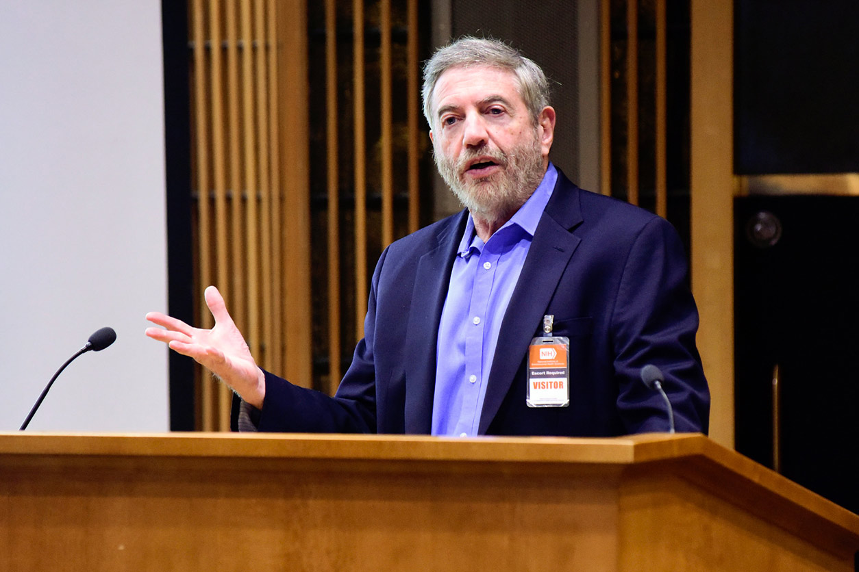
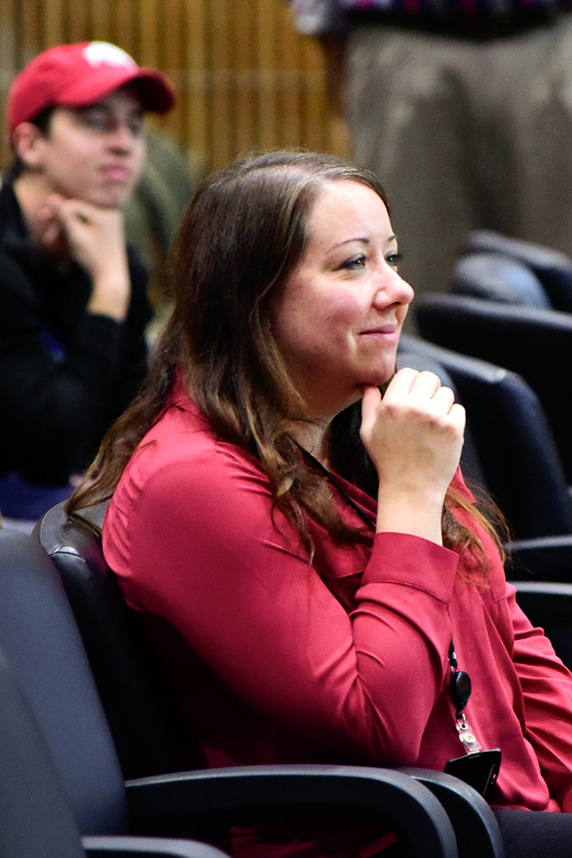
 Fessler, who introduced Tabas, was recently named head of the NIEHS Immunity, Inflammation, and Disease Laboratory. (Photo courtesy of Steve McCaw)
Mitochondria play a key role
Mitochondria in a resting macrophage are elongated in shape, which promotes interactions with another cellular organelle, called the endoplasmic reticulum (ER). This interaction enables a cell to isolate calcium within the two organelles.
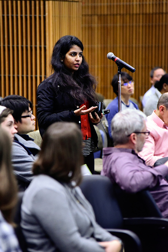 Chandana Kasireddy, Ph.D., a visiting fellow in the Integrative Bioinformatics Support Group, was one of the audience members who engaged Tabas with questions after his talk. (Photo courtesy of Steve McCaw)
When a macrophage encounters a dead cell, its mitochondria break up into smaller pieces, in a process known as fission. In this state, mitochondria and ER no longer interact, and calcium accumulates in the fluid of the cell.
Tabas explained that a gene called DRP1 regulates mitochondrial fission. The mitochondria of cells with defective DRP1 cannot carry out high-burden efferocytosis. He said this suggests the importance of mitochondrial fission in efferocytosis.
When macrophages with defective DRP1 genes cannot import calcium into their mitochondria, the cell’s calcium levels partially return to normal. Thus, said Tabas, calcium release into the fluid of the cell allows it to properly degrade dead cells and handle the restructuring of its own membrane.
Amino acid metabolism
A macrophage’s ability to internalize a second dead cell is coordinated with processing the metabolic load of the first dead cell, according to Tabas. So he reasoned that one or more of those metabolites would be involved in signaling to the macrophage to start internalizing a second dead cell.
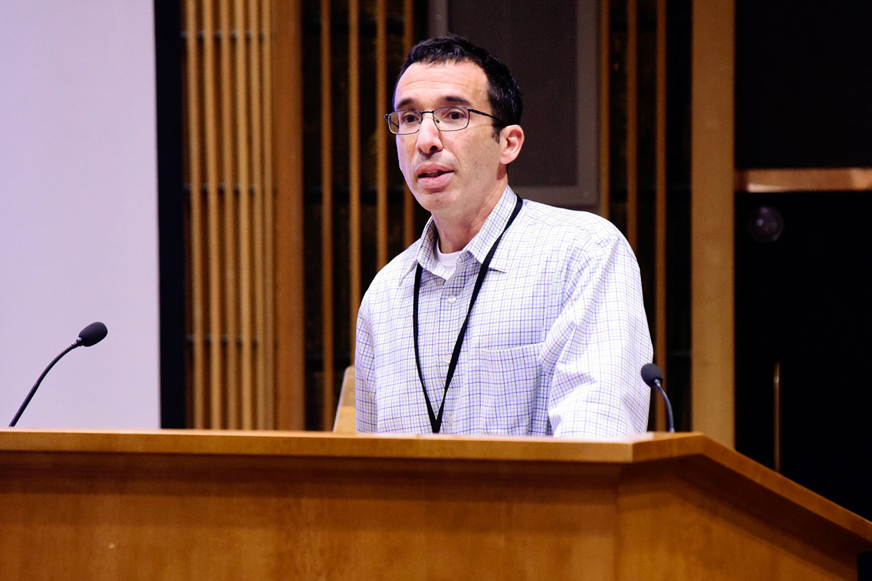
 Martinez’s research is related to that of Tabas’s, and during his talk, Tabas described how her work influenced his own. (Photo courtesy of Steve McCaw)
To learn more, he first sorted out macrophages that were and were not capable of internalizing two dead cells. Then he collaborated with Deb Muoio, Ph.D., from Duke University, to use a technique called liquid chromatography tandem mass spectrometry to show the cells that were able to perform efferocytosis.
The researchers identified significant changes in two amino acids — arginine and ornithine. By further studying cell strains that are deficient in converting arginine and ornithine, Tabas demonstrated a direct link between levels of the two amino acids and activation of high-burden efferocytosis.
“Tabas did a fantastic job of describing the coordinated dance that occurs between a dying cell and a phagocyte,” said Jennifer Martinez, Ph.D., head of the NIEHS Inflammation and Autoimmunity Group. “It’s not the simple act of clearing away debris, but a complex signaling relationship that requires participation from both parties — truly a ‘you are what you eat’ scenario.”
(Kiri Hoff, Ph.D., is an Intramural Research Training Award fellow in the NIEHS Mitochondrial DNA Replication Group.)
Citation: Wang Y, Subramanian M, Yurdagul Jr A, Barbosa-Lorenzi VC, Cai B, Juan-Sanz J, Ryan TA, Nomura M, Maxfield FR, Tabas I. 2017. Mitochondrial fission promotes the continued clearance of apoptotic cells by macrophages. Cell 171(2):331–345.e22.