A three-dimensional (3D) structure that allows scientists to see how and where disease mutations on the twinkle protein can lead to mitochondrial diseases has been developed by a team of scientists in the NIEHS Division of Intramural Research (DIR). Mitochondrial diseases are a group of inherited conditions that affect one in 5,000 people and have very few treatments.
The twinkle protein is involved in helping cells use energy converted from food. Prior to the development of this 3D structure, researchers were unable to determine how mutations contribute to disease.
“For the first time, we can map the mutations that are causing a number of these devastating diseases,” said lead author Amanda Riccio, Ph.D., a postdoctoral researcher in the Mitochondrial DNA Replication Group. “Clinicians can now see where these mutations lie, use this information to help pinpoint causes, and help families make choices, including decisions about having more children.”
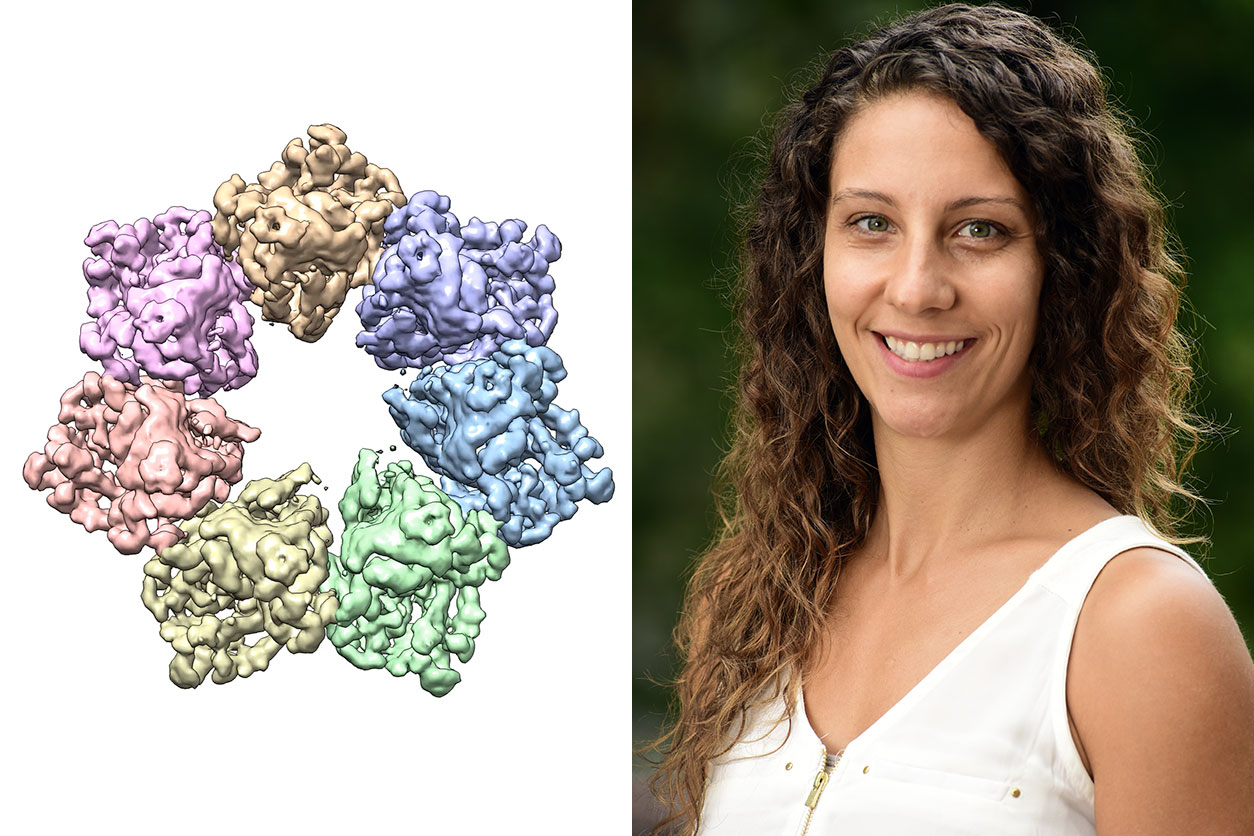 Riccio is the first to develop a 3D structure of the Twinkle protein. The structure enabled the research team to identify at least 25 mitochondrial disease-causing mutation sites within the protein structure. (Photos courtesy of Amanda Riccio and Steve McCaw / NIEHS)
Pathway to possibilities
The new findings will be particularly relevant for developing targeted treatments for patients who suffer from mitochondrial diseases such as progressive external ophthalmoplegia, a condition that can lead to loss of muscle functions involved in eye and eyelid movement; Perrault syndrome, a rare genetic disorder that can cause hearing loss; infantile-onset spinocerebellar ataxia, a hereditary neurological disorder; and hepatocerebral mitochondrial DNA (mtDNA) depletion syndrome, a hereditary disease that can lead to liver failure and neurological complications during infancy.
Published in the Proceedings of the National Academy of Sciences, the paper showcases how the NIEHS researchers were the first to accurately map clinically relevant variants in the twinkle helicase, the enzyme that unwinds the mitochondrial DNA double helix. The twinkle structure and all the coordinates are now available in the open data Protein Data Bank that is freely available to all researchers.
Final piece of mitochondrial puzzle
 Copeland has served as a longtime volunteer with the United Mitochondrial Disease Foundation. He is hopeful this breakthrough will lead to new treatments for such disease. (Photo courtesy of Steve McCaw / NIEHS)
“The structure of twinkle has eluded researchers for many years,” noted William Copeland, Ph.D., who leads the Mitochondrial DNA Replication Group and is the corresponding author on the paper. “It’s a very difficult protein to work with. By stabilizing the protein and using the best equipment in the world, we were able to build the last missing piece for the human mitochondrial DNA replisome.”
Using cryo-electron microscopy (Cryo-EM), the researchers were able to see inside the protein and the intricate structures of hundreds of amino acids or residues and how they interact.
Mitochondria, which are responsible for energy production, are especially vulnerable to mutations. mtDNA mutations can disrupt their ability to generate energy efficiently for the cell. Unlike other specialized structures in cells, mitochondria have their own DNA. In a cell’s nucleus, there are two copies of each chromosome. However, in mitochondria, there could be thousands of copies of mtDNA. Having a high number of mitochondrial chromosomes allows the cell to tolerate a few mutations, but accumulation of too many mutated copies leads to mitochondrial disease.
Precise location leads to precision science
To conduct the study, the researchers used a clinical mutation, W315L, known to cause progressive external ophthalmoplegia, to solve the structure. Using Cryo-EM, they were able to observe thousands of protein particles appearing in different orientations. The final structure shows a multi-protein, circular arrangement. They also used mass spectrometry to verify the structure and then performed computer simulations to understand why the mutation results in disease.
Within twinkle, they were able to map up to 25 disease-causing mutations. They found that many of these disease mutations map right at the junction of two protein subunits, suggesting that mutations in this region would weaken how the subunits interact and make the helicase unable to function.
 Longley, a scientist in Copeland’s group, is shown here delivering a lecture at Duke University in 2018 as part of the University Program in Environmental Health and Toxicology Seminar Series. (Photo courtesy of Steve McCaw / NIEHS)
“The arrangement of twinkle is a lot like a puzzle,” Riccio explained. “A clinical mutation can change the shape of the twinkle pieces, and they may no longer fit together properly to carry out the intended function.”
Paving way for treatments
“What is so beautiful about Dr. Riccio and the team’s work is that the structure allows you to see so many of these disease mutations assembled in one place,” said Matthew Longley, Ph.D., an author on the paper and NIEHS associate scientist.
“It is very unusual to see one paper that explains so many clinical mutations,” Longley said. “Because of NIEHS’ investments in state-of-the art Cryo-EM equipment and talented people like Amanda Riccio who have the skill set to work on this challenging protein, we are one step closer to having information that can be used to develop treatments for these debilitating diseases.”
Longley’s sentiments were echoed by Philip Yeske, Ph.D., from the United Mitochondrial Disease Foundation (UMDF).
“Congratulations are in order for the team at NIEHS for this groundbreaking work on twinkle that ultimately may lead to treatments and cures for mitochondrial disease patients,” said Yeske, who serves as the UMDF Science and Alliance Officer. “Our community is fortunate to have such a talented group of scientists conducting basic research to understand the biological underpinnings of mitochondrial disease, and I look forward to future translational work in this area.”
Citation: Riccio AA, Bouvette J, Perera L, Longley MJ, Krahn JM, Williams JG, Dutcher R, Borgnia MJ, Copeland WC. 2022. Structural insight and characterization of human Twinkle helicase in mitochondrial disease. PNAS (119)32 e2207459119.
(Robin Mackar is a writer and media relations coordinator in the NIEHS Office of Communications and Public Liaison.)