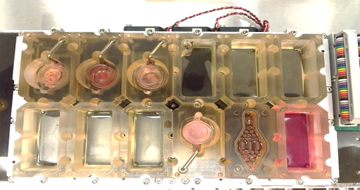
Shuo Xiao, Ph.D., recently developed a miniature model to more accurately study how environmental exposures affect infertility and women’s reproductive health. The 3D models, developed in part through a series of NIEHS grants, mimic the female reproductive tract.
The “organs-on-a-chip” device contains cells from four reproductive organs — the ovaries, fallopian tubes, uterus, and cervix — that grow into organoids, a 3D structure that mimics the function of an organ. These organoids are connected by a series of small tubes that carry hormones and other signals between them.
“The miniature chip replicates the hormone fluctuations and cellular changes that occur over a woman’s menstrual cycle,” explained Xiao. “When we connect a mini 3D liver to the device, we can replicate how the reproductive system reacts to a drug or toxin after it has been metabolized by the liver.”
Expanding beyond endocrine disruptors
The current models build on Xiao’s previous research investigating the effects of endocrine-disrupting chemicals. Working under the guidance of Xiaoqin Ye, Ph.D., Xiao received his doctorate in female reproductive toxicology at the University of Georgia. There, he used mouse models to study how exposure to endocrine-disrupting chemicals affect embryo implantation in the uterus, a critical step for a successful pregnancy.
For his postdoctoral research, he continued investigating how the environment affects women’s reproductive health, with two major differences. He expanded his studies beyond the uterus to incorporate other organs in the female reproductive system and sought to reduce the use of animal models in his research.

Fine tuning for toxicology
Xiao recognized the immense potential of the chip platform for chemical toxicity testing; however, the original method needed some tweaking.
“With the initial device, it could take months to test a chemical at different doses to determine its toxicity," said Xiao. "I wanted to optimize the approach so we could use the device to rapidly screen a broad range of chemicals.”
With an NIEHS career development award, Xiao moved to the University of South Carolina to focus on fine-tuning one component of the device for toxicity testing: the follicle. The follicle is a small sac in the ovary that contains an egg and produces hormones crucial for ovulation and fertilization. Environmental exposures that alter follicle hormone secretion or egg quality may reduce a woman’s fertility.
Under the mentorship of Jodi Flaws, Ph.D., and Mary Zelinski, Ph.D., Xiao developed a method, called vitrification, to freeze immature follicles so they can be stored long-term, thus creating a ready-to-use ovarian follicle bank for high-throughput toxicity screening. He demonstrated that, when thawed, preserved cells behaved on par with fresh follicles. Specifically, warmed follicles grown in the “ovary-on-a-chip” environment developed into 3D organoids, released reproductive hormones, initiated ovulation, produced a mature egg cell, and had similar expression of genes important in these processes.
Taking inspiration from his postdoctoral work and with funding from a second NEIHS grant, Xiao is now connecting a liver organoid to the ovary-on-a-chip. This addition will better mimic how follicles respond to toxins in the body that have been transformed by the liver.
(Megan Avakian is a senior communication specialist at MDB, Inc., a contractor for the NIEHS Division of Extramural Research and Training.)


