The complex relationship between environmental stressors, extracellular vesicles (EVs), and adverse health outcomes was the focus of an NIEHS workshop held Sept. 27-28.
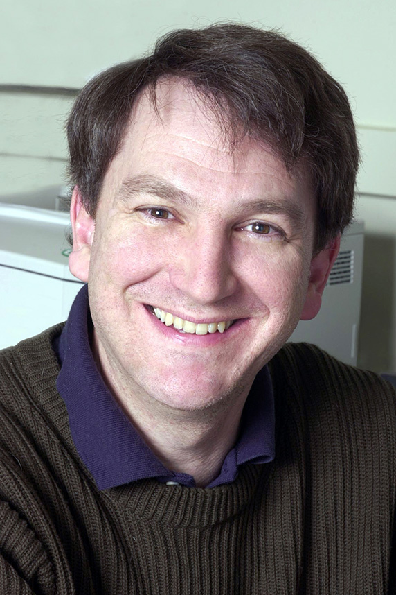 Shaughnessy manages grants on the development and validation of biomarkers of response to environmental stress, among his other duties at NIEHS. (Photo courtesy of Steve McCaw / NIEHS)
EVs shuttle proteins, nucleic acids, and lipids to cells, and they play a key role in cell-to-cell communication. They have been linked to a wide range of biological activity involving both normal and disease states. Examples include immune responses, repair of injured tissue, and anti-tumor and pro-tumor processes.
Understanding EV function may help researchers identify early biological signs of the effects of environmental exposures and shed light on disease progression, according to NIEHS Health Scientist Administrator Daniel Shaughnessy, Ph.D., who helped to organize the event.
Identifying knowledge gaps, opportunities
Workshop goals included the following.
- Discuss the state of the science and technology related to how environmental stress affects EVs and cell signaling.
- Identify both knowledge gaps and opportunities for integrating EV analysis into environmental health sciences research.
Stephen Gould, Ph.D., from Johns Hopkins Medicine, described the biology of EV formation and release by cells, and he shared some of the challenges of detecting the vesicles in blood and other biospecimens. Several participants, including NIEHS grantees, explored how to analyze EVs to assess biological signs of injury following environmental exposures.
Andrea Baccarelli, M.D., Ph.D., from Columbia University, talked about his research on the role of EVs generated in the lung in response to air pollution exposure. He noted potential effects on lung function and inflammation that can affect other organs, such as the brain.
Other scientists discussed how the environment can affect EVs and influence conditions such as Parkinson’s disease and amyotrophic lateral sclerosis, or ALS.
EVs — emerging area of interest
 Jones has established a translational EV pipeline that includes instrumentation and tools for analysis, calibration, and benchmarking efforts. (Photo courtesy of Peter Guion / NCI)
“There is so much interest in this exciting, emerging field,” said Jennifer Jones, M.D., Ph.D., a Stadtman Investigator at the National Cancer Institute (NCI). “I have noticed more and more early-career scientists enthusiastically embrace the potential for EVs to inform their research.”
Exosomes — a subset of EVs — are of particular interest. They carry messenger RNA, proteins, and other molecules to aid signaling between cells during normal conditions but also during stress resulting from exposure to toxicants. Such movement of molecules can affect distant cells and tissues, potentially leading to biological injury and disease.
“If exosomes represent early indicators of injury, they may provide us with a means of studying pathogenesis to a point where interventions may be more practical and easier to implement,” noted Kenneth Ramos, M.D., Ph.D., from Texas A&M University.
Ramkumar Menon, Ph.D., from the University of Texas Medical Branch at Galveston, described his work analyzing exosomes in the context of premature birth. He used an organ-on-chip device that mimics human biological interactions between fetus and mother. Menon showed that fetal exosomes shuttling the protein HMGBI functioned as a signal to trigger early labor.
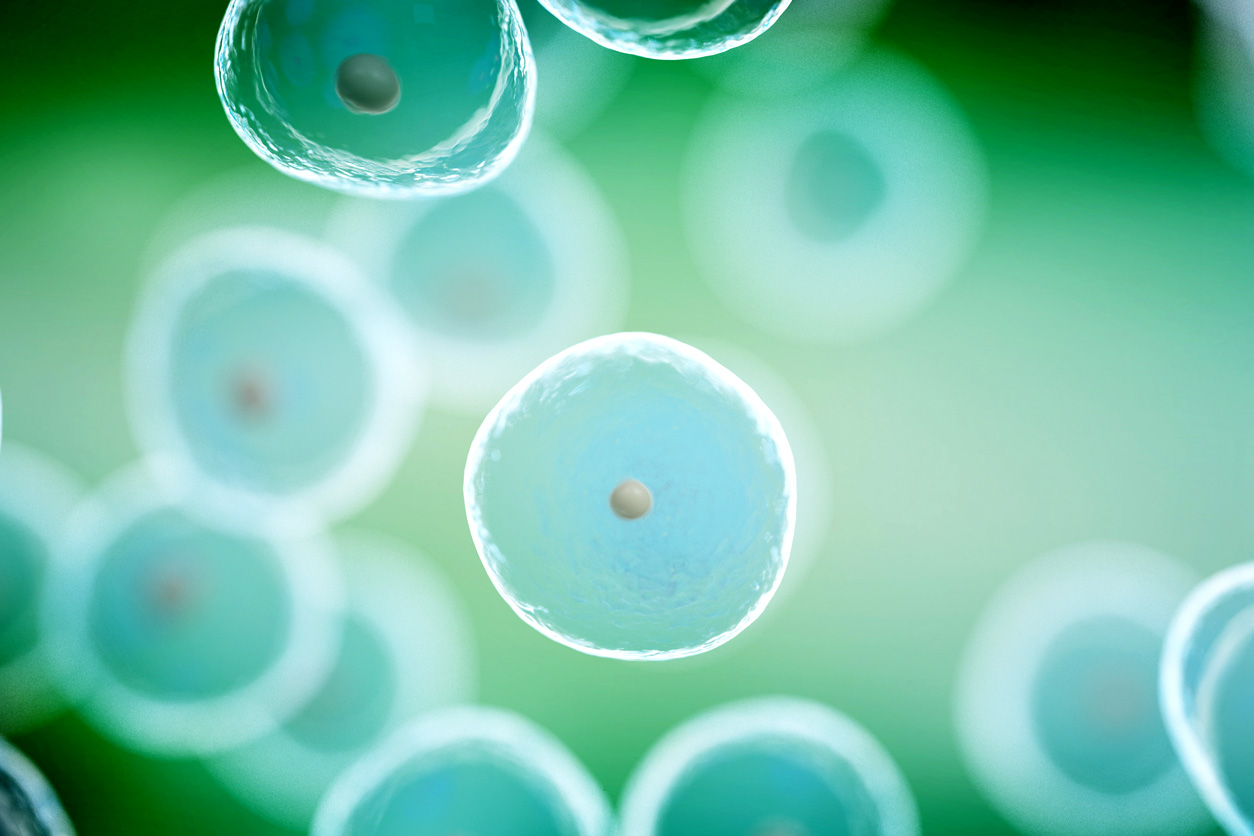 EVs are a key means of communication between cells in the same tissue and between organs, and they encompass a wide range of sizes and biological functions. (Photo courtesy of SciePro / Shutterstock.com)
Only scratching the surface
NIEHS scientists Alex Merrick, Ph.D., and Ann Jukic, Ph.D., moderated forums in which participants shared current knowledge on EVs and discussed scientific gaps. Many attendees expressed the need for further research in this important area.
“The biggest challenge in terms of studying the exosome is on the methods side,” said Merrie Mosedale, Ph.D., from the University of North Carolina at Chapel Hill. “We would like a uniform approach that can be applied in toxicology studies, but we are not there yet.”
“If you look at where we are in the field right now, especially in the presentations we heard over the last two days, we are only beginning to scratch the surface,” said Ramos.
Citations:
Doyle LM, Wang MZ. 2019. Overview of extracellular vesicles, their origin, composition, purpose, and methods for exosome isolation and analysis. Cells 8(7):727.
Radnaa E, Richardson LS, Sheller-Miller S, Baljinnyam T, Silva M, Kammala AK, Urrabaz-Garza R, Kechichian T, Kim S, Han A, Menon R. 2021. Extracellular vesicle mediated feto-maternal HMGB1 signaling induces preterm birth. Lab Chip 21(10):1956–1973.
(Tara Ann Cartwright, Ph.D., is a technical writer-editor in the NIEHS Office of Communications and Public Liaison.)


