A new exhibit of scientific images made by NIEHS researchers using cutting-edge technology is being hung throughout the institute. As the pandemic eases in the coming months and more employees return to work onsite, they will be greeted by 27 new framed images displayed in laboratory and administrative areas.
Organized by a group of NIEHS trainees led by Joseph Dahl, Ph.D., former president of the NIEHS Trainees’ Assembly (NTA), the images were selected through a competition launched by Scientific Director Darryl Zeldin, M.D. “It has been over five years since our last collection of images,” said Zeldin, referring to an earlier display.
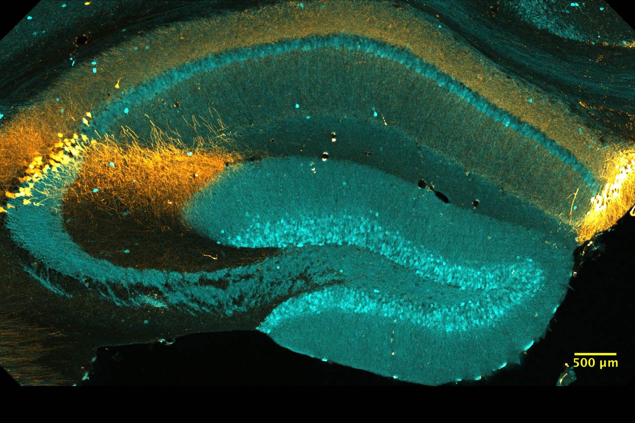 Coronal section of a mouse brain hippocampal area CA2 expressing a fluorescent protein under the control of the Amigo2promoter (yellow). CA2 neurons are molecularly distinct from the neighboring hippocampal CA subfields. By Shannon Farris, Ph.D., and Benjamin Slay, working with Serena Dudek, Ph.D. (Image courtesy of NIEHS)
Cross-section and time capsule
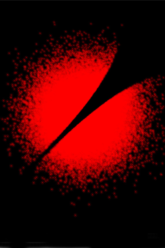 Decorrelated statistics plotted against each other show a gap in the circle, which illustrates that a lack of correlation does not imply independence. By Dmitri Zaykin, Ph.D. (Image courtesy of NIEHS)
Dahl explained that the selections represent branches and labs from across the institute, including the Clinical Research Unit and the Division of the National Toxicology Program.
“Environmental health sciences encompass so many fields — from genomics to cloning to the way that drugs affect cells and tissue,” he said. “The collection represents both a cross section of that work, and a time capsule of techniques in use today.”
Dahl remains enthusiastic about the months-long project. “Dr. Zeldin is such a great advocate for the fellows, so when he asked me to do this, I tried to make it happen,” Dahl said.
Emphasis on education
Joining Dahl on the NTA committee were Nancy Urbano, Kiana Gunn, and Thanh Hoang, Ph.D. They discussed selections with both leadership and interior design teams. The educational aspect was a high priority, showing how tissue responds to external stimulus and fine structural detail.
Framed photographs will be hung in various hallways and lobbies, for viewing by visitors who tour the institute. “We want to both educate viewers and showcase the amazing work being done here,” Dahl explained. “It is also a great way to demonstrate what our arts and photography team can do.”
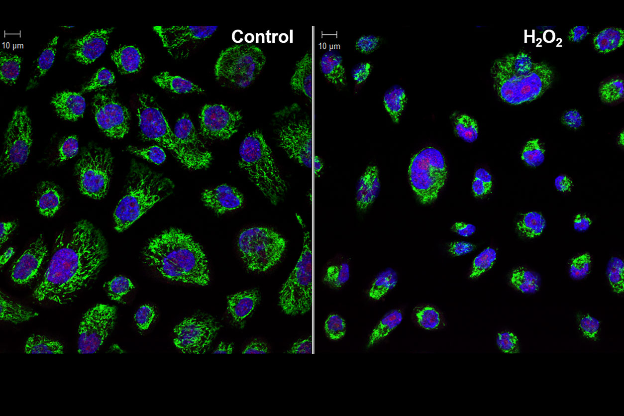 Hydrogen peroxide damages mitochondria, as shown by reduced Tom20 staining (green) in primary immortalized human bronchial epithelial cells. Blue: nuclei. Red: human DNA glycosylase NTHL1. By Aniraban Kar, Ph.D., working with Paul Doetsch, Ph.D. (Image courtesy of NIEHS)
Dahl thanked the Office of the Scientific Director, the trainees, and especially the individuals who did the science that is represented.
NIH collects for public sharing
The final collection was sent to the National Institutes of Health (NIH) in response to a call by the NIH Office of Intramural Research (OIR). “We intend to use these images both on the IRP [Intramural Research Program] web site and on our social media platforms as a visual hook to entice people to click through and learn more about our research accomplishments,” wrote Andy Baxevanis, Ph.D., OIR director of computational biology, in his email request.
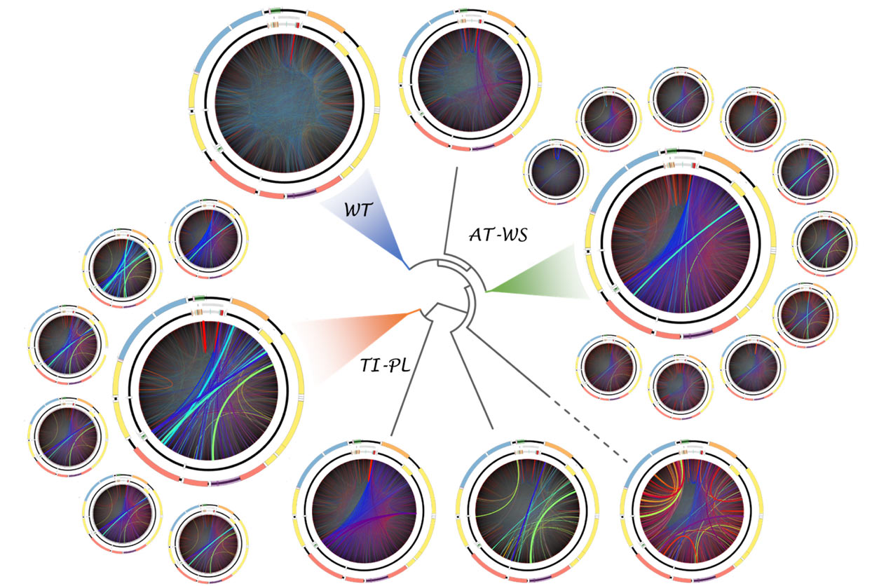 Deletions in mitochondrial DNA, shown by colored arcs, distinguish normal human skeletal muscles (WT) from those with POLG-driven disease, revealing processes correlated with aging and DNA replication mechanisms. By Scott Lujan, Ph.D., working with Bill Copeland, Ph.D., and Tom Kunkel, Ph.D. (Image courtesy of NIEHS)
“Intramural scientists are constantly producing amazing images that inform and advance scientific progress,” wrote Baxevanis. “As an added bonus, they also provide us a phenomenal way of catching the public’s eye and educating them about the cutting-edge work being done in the IRP.”
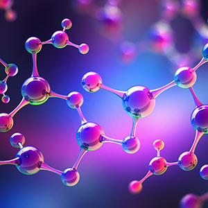
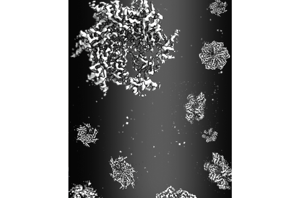 Cryo-electron microscopy snapshots of Rix7, an essential compound in ribosome production, revealed its function as an asymmetric hexamer that unravels substrates through its central pore. By Yu-Hua Lo, Ph.D., working with Robin Stanley, Ph.D. (Image courtesy of NIEHS)
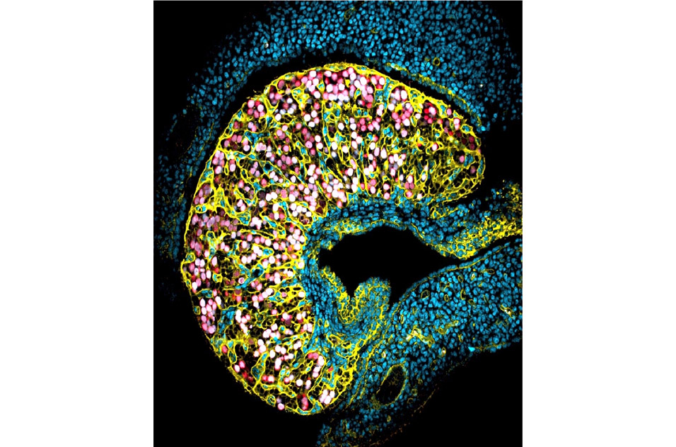 In a mouse fetal ovary, future oocytes (red and white) are enclosed in nests by a basal membrane (yellow) that separates them from the interstitial cells (blue). By Barbara Nicol, Ph.D., working with Humphrey Yao, Ph.D. (Image courtesy of NIEHS)
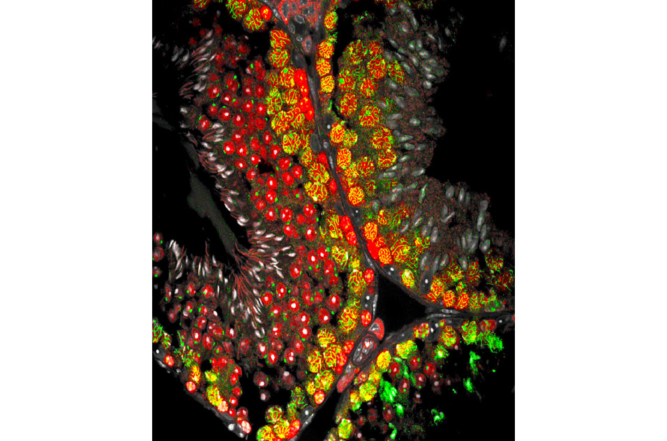 In an adult mouse testis, seminiferous tubules contain germ cells (red) that differentiate into spermatozoa (white). Green lines highlight pairing of homologous chromosomes in meiotic spermatocytes. By Barbara Nicol, Ph.D., working with Humphrey Yao, Ph.D. (Image courtesy of NIEHS)
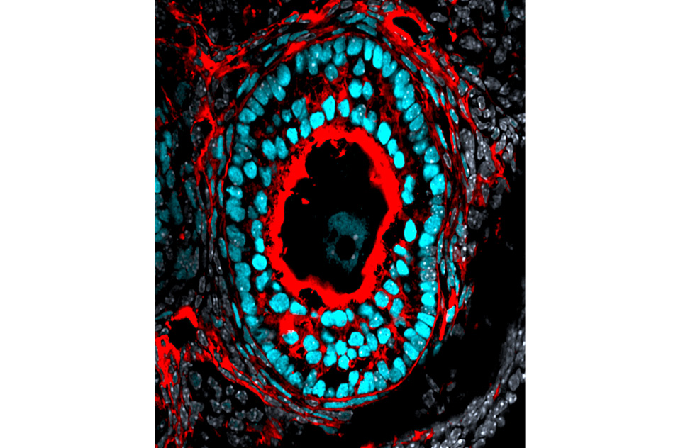 Several cell types are found in the adult ovary preantral follicle. Cyan: granulosa cells. Red: interstitial cells. Gray: other nuclei. By Saniya Rattan, Ph.D., working with Humphrey Yao, Ph.D. (Image courtesy of NIEHS)
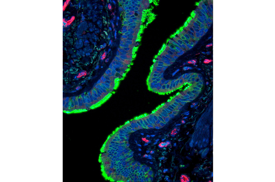 Cilia expression (green) on epithelial cells (nuclei in blue) line the airway in a human lung section. By Carol Trempus, working with Stavros Garantziotis, M.D. (Image courtesy of NIEHS)
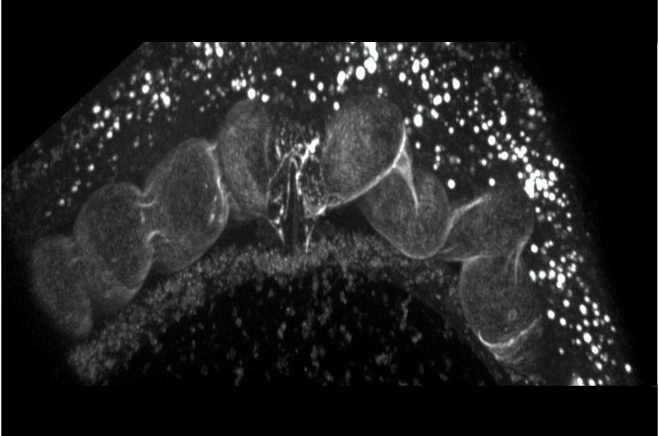 Confocal microscopy of a live Caenorhabditis elegans worm shows embryos in utero. By Sandra Vergara, Ph.D., working with Perry Blackshear, M.D., D.Phil. (Image courtesy of NIEHS)
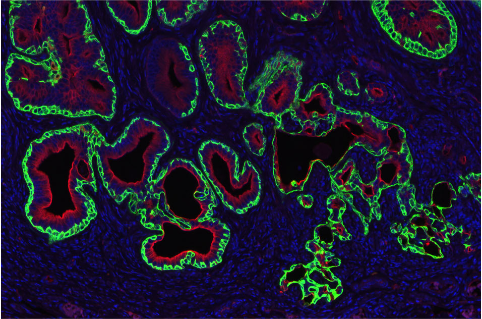 Endometrial carcinoma in an adult mouse exposed neonatally to diethylstilbestrol. Green: squamobasal cells. Red: glandular epithelial cells. Blue: epithelial and stromal nuclei. By Alisa Suen Wallach, Ph.D., working with Carmen Williams, M.D., Ph.D. (Image courtesy of NIEHS)
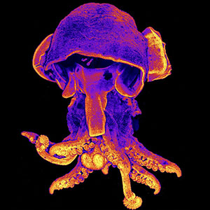
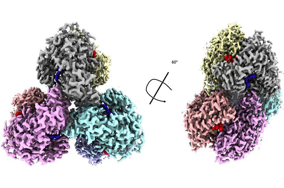 Monomers of the hexameric protein dGTPase from the bacterium Leeuwenhoekiella blandensis are individually colored in this cryo-electron microscopy image. The protein is bound to the dGTP substrate (blue) and dATP (red), an allosteric activator. By Brad Klemm, Ph.D., working with Roel Schaaper, Ph.D. (Image courtesy of NIEHS)
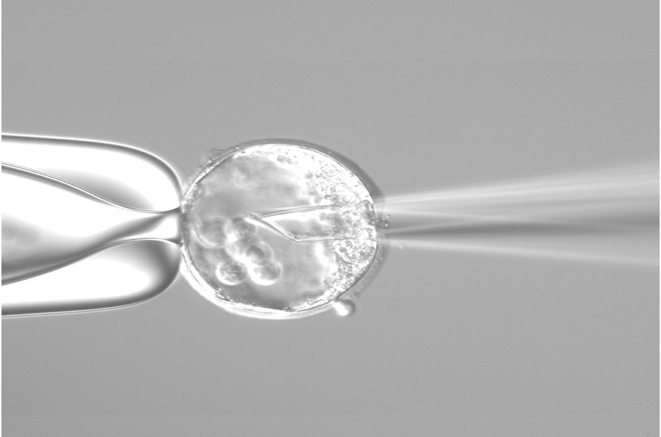 This image shows a stage in the creation of a genetically modified mouse at NIEHS. By Gregory Scott, working with Manas Ray, Ph.D. (Image courtesy of NIEHS)