When “The Beautiful Brain” opened Jan. 25 at the University of North Carolina at Chapel Hill (UNC), the show of anatomical drawings by Spanish artist and Nobel-winning scientist Santiago Ramon y Cajal included a scientific image from the lab of Serena Dudek, Ph.D., deputy chief of the NIEHS Neurobiology Laboratory.
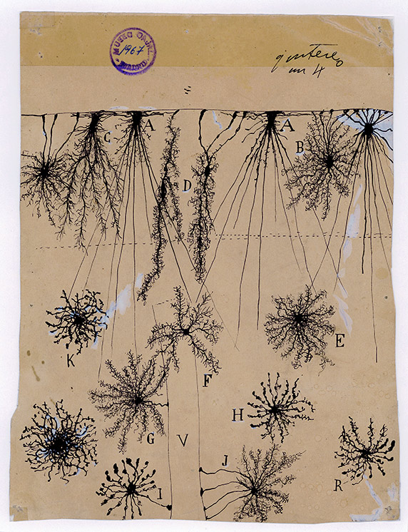 Drawing by Santiago Ramón y Cajal of glial cells of the cerebral cortex of a child. Made in 1904, ink and pencil on paper. (Image courtesy of Instituto Cajal)
Cajal, known as the father of neuroscience, trained first as an artist. “I think all neuroscientists appreciate his contributions,” said Dudek, noting that his drawings are still used in teaching today.
Cajal was a contemporary of Camillo Golgi, with whom he shared a Nobel Prize in 1906. Yet they held contrary views of the brain’s structure. “Cajal used Golgi’s methods, but the two were infamous for their rivalry,” said Dudek, who also serves on the neuroscience faculty at the UNC School of Medicine.
“Cajal offered what is now known as the neuron doctrine, which holds that neurons are separate entities,” she explained. “Golgi thought they were all physically part of the same network or structure.” She added that the question was not resolved definitively until the 1950s, with the advent of electron microscopy.
Imaging leads to unexpected findings
The NIEHS image on display at UNC’s Ackland Art Museum is from a remarkable study published in 2016. The dark areas in the electron micrograph show structures called perineuronal nets (PNNs) in a surprising location, according to lead author Kelly Carstens, Ph.D., a postdoctoral fellow in Dudek’s lab. That location is in a part of the brain’s hippocampus known as CA2, which is involved in social behaviors, learning, and memory.
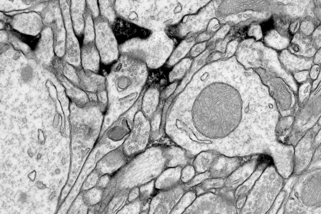 “Our black stain [PNNs] encompassed synapses around pyramidal neurons,” Carstens explained, pointing to the mushroom shapes, center and lower right. “This leads to questions about whether PNNs have an important function in regulating synaptic plasticity … and ultimately learning and memory. (Image courtesy of Carstens, et al.)
Dudek is a pioneer in the study of CA2 development. “We were the first to find that in hippocampal area CA2, PNNs surround dendritic spines on pyramidal neurons, which are excitatory,” she explained. Excitatory neurons stimulate further signaling. PNNs were previously known to surround inhibitory neurons, so named because they slow or shut down signals.
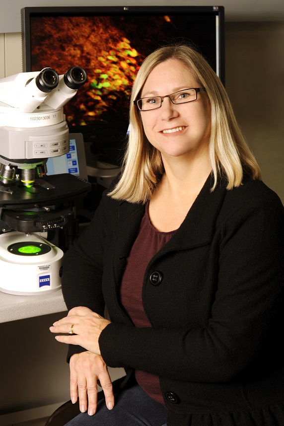 Dudek serves as deputy chief of the NIEHS Neurobiology Lab and also leads the Synaptic and Developmental Plasticity Group. (Photo courtesy of Steve McCaw)
Carstens and the rest of the team believe their findings have important implications for understanding a critical early period of development. “Developmental regulation of PNNs in area CA2 may … represent a therapeutic target for PNN-associated developmental disorders, such as schizophrenia and Rett syndrome,” they wrote.
Carstens carried out the research while a predoctoral fellow in Dudek's lab and a graduate student in the UNC lab of Richard Weinberg, Ph.D. He is a co-author on the paper.
Art may understand before science proves
Although Cajal thought PNNs were merely a result of tissue preparation and staining, his drawings depicted them meticulously. “I’m always humbled by how much there is still to learn from drawings of brain anatomy that were done over 100 years ago,” Carstens said. “The brain area Serena studies was overlooked for many decades, but it was actually described as a distinct region of the hippocampus in 100-year-old drawings.”
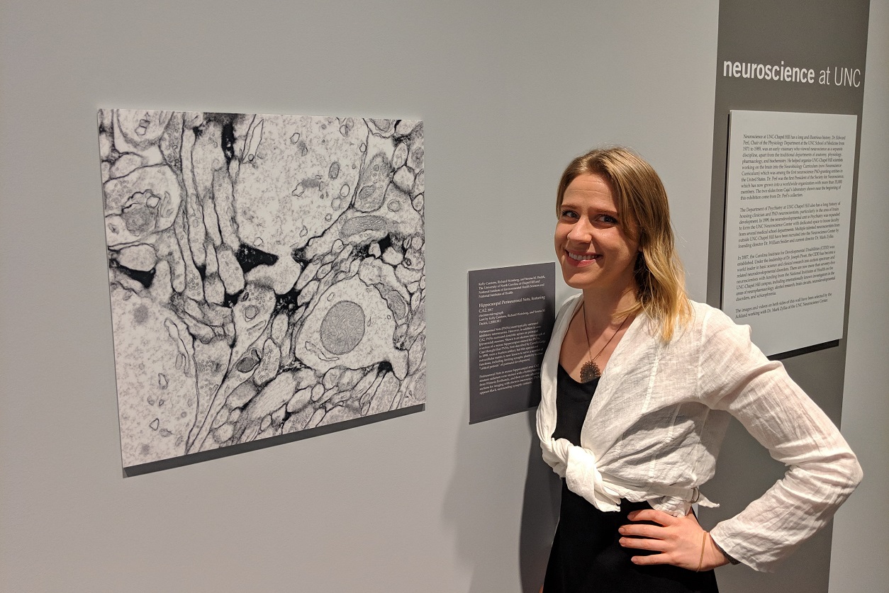 “We wanted to determine the location more specifically in the brain to give us clues about its potential function in the brain,” Carstens said, shown at the exhibit. (Photo courtesy of Steve McCaw)
“Drawing was a tool to observe, discern, and recount microanatomy structure,” wrote artist Dawn Hunter, in a November 2017 blog post for the National Library of Medicine (NLM). Several Cajal drawings are on display at the National Institutes of Health John Edward Porter Neuroscience Research Center. A 2017 Fulbright Scholarship allowed Hunter to Cajal’s study sketchbooks and other materials at the Instituto Cajal in Madrid.
She gained deep insights into the connection between drawing and scientific understanding, as reported in a follow-up NLM blog post in October 2018. “He does not erase or re-draw his lines,” she observed. “The only times when his lines demonstrate hesitation or confusion are on the pages where he drew neurons for the first time…. I have surmised that the inconsistency is because of an incorrect expectation on his part; he thought from the onset that those lines would interconnect or meet.”
The act of drawing appears to have shown him that the brain differed from Golgi’s understanding and may have helped him develop the neuron doctrine.
Citations:
Carstens KE, Phillips ML, Pozzo-Miller L, Weinberg RJ, Dudek SM. 2016. Perineuronal nets suppress plasticity of excitatory synapses on CA2 pyramidal neurons. J Neurosci 36(23):6312–6320.
Vitellaro-Zuccarello L, De Biasi S, Spreafico R.1998. One hundred years of Golgi's 'perineuronal net': history of a denied structure. Ital J Neurol Sci 19(4):249–253.